
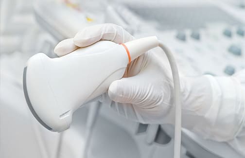
Ultrasound is a painless and harmless examination of the thyroid gland, and along with a clinical examination and determination of thyroid hormones, it must be one of the first diagnostic examinations of the thyroid gland. With it, you can clearly see the size and structure of the left and right lobes and the middle part of the thyroid gland, which is called the isthmus. From this, we can calculate the weight and volume of the thyroid gland, which is particularly important when monitoring the size of a diffusely enlarged thyroid gland. Also, chronic inflammation of the thyroid gland, which is called Hashimoto's thyroiditis, gives a very characteristic ultrasound picture.
However, ultrasound is of particular importance in monitoring the size, limitation and structure of one or more nodes in the thyroid gland, and calcifications can also be seen. Also, ultrasound can find nodes that are larger but are located at the back, as well as smaller ones, only a few millimeters in size, which are not palpable and can already be cancer with metastases on the neck. Nodes should be punctured under ultrasound control as soon as they are found. Determine the exact position of the tip of the needle. A possible thyroid cancer is ruled out by a puncture. Ultrasound examination of the neck in nodular thyroid should be repeated every 6 to 12 months. and in the case of an increase in the size of a node, it is recommended to repeat the cytopuncture. In addition, ultrasound can be used to monitor neck lymph nodes as well as the appearance of new lymph nodes, which is particularly important after thyroid cancer surgery.
Ultrasonographic examination of the thyroid is of particular importance in the early detection of thyroid cancer, which is often accidentally detected by ultrasound. The sooner we detect it, the better the prognosis for patients, as in all other cancers.
No special preparation is required, the examination itself takes about 15 minutes, but with a conversation with the patient and writing the findings, it can take about 40 minutes.


Comments are closed.