What is ERCP?
Endoscopic retrograde cholangiopancreatography (ERCP) is a procedure that combines upper gastrointestinal (GI) endoscopy and x-rays to treat problems with the bile and pancreatic ducts.
When is ERCP performed?
Doctors perform an ERCP when your bile ducts or pancreatic ducts have become narrowed or blocked because of:
- Gallstones (formed in the gallbladder) in the common bile duct,
- infection,
- acute and chronic pancreatitis
- trauma or surgical complications in your bile or pancreatic ducts
- pancreatic pseudocysts
- bile duct tumors or cancers
- tumors or cancers of the pancreas
ERCP is primarily used to diagnose and treat conditions of the bile ducts and main pancreatic duct. ERCP can be performed for diagnostic and therapeutic reasons, although the development of safer and relatively non-invasive investigations such as magnetic resonance cholangiopancreatography (MRCP) and endoscopic ultrasound have meant that ERCP is now rarely performed without a therapeutic intent.
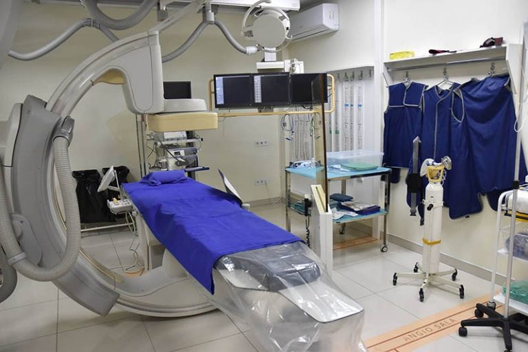
How is ERCP performed?
An intravenous (IV) needle will be inserted into your arm to give you a sedative. A sedative will be injected, and you will feel relaxed and sleepy almost immediately. While you are under sedation, your vital functions are continuously monitored (the amount of oxygen in your blood and the values of your blood pressure and pulse). You will be asked to lie down on the examination table. The doctor will carefully guide the duodenoscope down your esophagus, through your stomach, and into your duodenum. A small camera placed on the duodenoscope sends a video image to a monitor. A duodenoscope pumps air into your stomach and duodenum, making them easier to see. During ERCP, the doctor:
- locates the opening where the bile and pancreatic ducts drain into the duodenum,
- passes a thin, flexible tube called a catheter through the duodenoscope into the ducts
- injects a special dye, also called a contrast agent, to make the canals more visible on X-rays
- uses a type of x-ray, called fluoroscopy, to examine the ducts and look for narrowed areas or blockages
The doctor can pass small tools through the duodenoscope to
- opened blocked or narrowed ducts.
- broke or removed stones
- performed a biopsy or removed tumors in the ducts.
- inserted stents—tiny tubes that doctors leave in narrowed ducts to keep them open.
The procedure usually lasts between 1 and 2 hours.
We are the first in the region to perform insufflation with CO2
Advantages of insufflation with CO2 compared to atmospheric air